

Introduction: Perspectives on Size
Visible thinking routine: See-think-connect. Watch the video on size.
- Acting as a class, summarize everything that you saw (from memory in the video)
- Harvest the thoughts from the class that members have about what they have seen in the video
- Connect these thoughts to concepts you have learned in other subjects, or thoughts you have had outside of the classroom.
Lesson one: A2.2.1-A2.2.5
Key objectives:
1) Learn how to look at cells (microscope skills)
2) Learn how to make cells (wet mount)
3) Learn how to calculate scale
4) Learn how to draw a detailed bacterial cell (prokaryotes)
Key vocabulary:
- Cells: the smallest unit of life.
- Scale: The estimate of how big an image is relative to the real size
- Micrometer (one thousandth of a millimetre)
- Nanometer (one millionth of a millimetre)
- Light microscope: A device for viewing small things that uses light.
- Electron microscope: A device for viewing small things that uses electrons, and allows for magnified images with greater resolution.
- Prokaryotes: A kind of cell that includes true bacteria and other bacteria.
A2.2.1 Cells are the basic structural unit of all living organisms.
Cell theory states that all living things are made from cells. The theory was developed by the first biologists to view living tissues using a microscope, including the inventor of the modern light microscope, Robert Hooke.
Although exceptions have been found to this theory, it is still useful and has not been rejected.
A2.2.2 Microscope skills
The human eye can perceive an object as small as 0.1mm, but smaller objects cannot be detected without the help of a microscope.
A microscope with an eyepiece of x10 and a high power lens of x 40 will magnify an object by x400. This will allow humans to perceive an objects which are 400 times too small to see with the naked eye.
Before completing the microscope lab, practice the parts of a light microscope
A2.2.3 Developments in microscopy
Light microscopes can be used to view living tissues, without harming them. Light microscopes also can be used to view tissues in color, although chemical stains (for example methylene blue) are often used to enhance colors to make certain components more visible. Light microscopes can only magnify by x400, without images becoming blurry, or low resolution.
Electron microscopes can be used to create high resolution images of living tissues at much greater magnifications. Electron microscopy always kills the tissues.
While electron microscopes are more powerful, both kinds of microscopes are useful in modern biology.
Q) Suggest a context in which it might be better to use a light microscope over an electron microscope, and vice versa. Justify your statement.
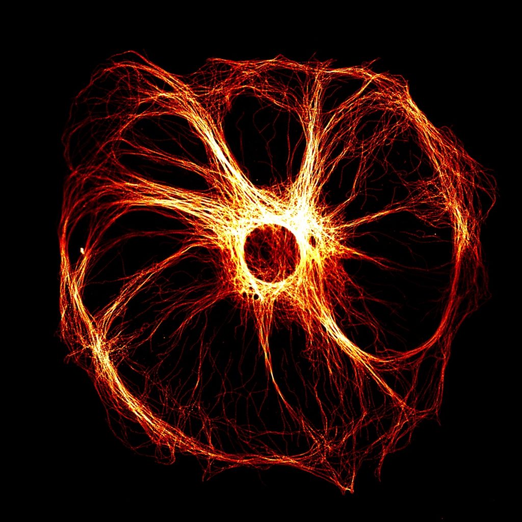
The image above was captured by Derek Sung in 2017, and captures a Sertoli cell, a kind of cell in the human testes.
A technique known as immunoflourescence was used to create this image.
Simply stated, antibodies are chemicals made by white blood cells that are designed to attach to certain chemicals called antigens. These antibodies can be combined with flourescent markers. The result may be a beautiful image such as this one, where antibodies are binding to specific proteins and causing parts of an image to flouresce.
Freeze-fracture electron microscopy is a technique used to produce detailed images of cell surfaces, showing the three dimensional aspect of organelles.
Stages of freeze-fracture electron microscopy:
- Sample is plunged into liquid nitrogen, freezing the contents
- Cell is split with a steel blade, along the weakest point
- Some of the ice is vaporised at the surface, to clarify the form (etching)
- The surface is coated with vaporised platinum or carbon, which freezes to form a coating in the exact shape of the surface.
Even cell membranes can be split using freeze-fracture microscopy, this led to the discovery that the cell membrane had proteins embedded in it.
Activity20 min: Visit the scanning electron microscope simulator SEMsimulator Learn how to adjust the settings, and save one high quality image you can share with the class.
Cryogenic electron microscopy Is similar to freeze fracture in that the sample is frozen. In this case, to -180 oC. However cryogenic electron microscopy is usually used to examine protein structure, and so the sample is often a solution containing proteins. Freezing the sample makes the proteins more stable.
Activity 5min: Glossary charades. Using hand gestures and body movements, choose either cryogenic electron microscopy, freeze-fracture electron microscopy or immunoflourescence and see if your peers can guess the key term you are illustrating.
A2.2.4 Structures common to cells in all living organisms.
Activity: Test your pre-knowledge. Sketch a cell of any kind and label the plasma membrane, cytoplasm, and the DNA.
The plasma membrane is the perimeter of the cell, and it controls the passage of substances into and out of the cell.
The cytoplasm comprises the main content of the cell and consists of a solution of proteins, ions and many other substances suspended or dissolved in water.
The DNA contains the genetic information required to make specific proteins, control metabolic reactions, and to replicate the cell.
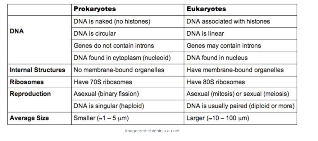
A2.2.5 Prokaryotic cell structure
All cells can be divided into two main types:
- Those with a nucleus (this includes the cells of aninals, plants, fungi and most organisms). The term eukaryotic is used to refer to these types of cells.
- Those without a nucleus (this includes bacteria only). The term prokaryotic is used to refer to these types of cells.
Refer to the image of a prokaryotic cell, belonging to the bacterium Clostridium botulinim.
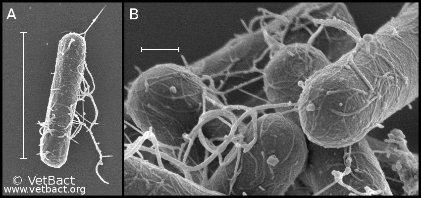
Scale bars A and B represent the distances 4 and 0.4 micrometres respectively.
Activity: Calculate the magnification of the image and describe the shape.
Prokaryotes such as Clostridium have a cell wall, which is a barrier outside of the cell membrane to protect the cell. This is made of a substance called peptidoglycan.
The cells of prokaryotes are filled with cytoplasm, and not subdivided into different compartments by internal membranes.
They will contain structures called ribosomes (smaller size 70S) where they manufacture proteins.
Their DNA consists of a single DNA loop, without proteins (naked). The part containing the DNA is called the nucleoid, but it is not a nucleus.
Prokaryotes may have long thread like proteins protruding from their surfaces, these may be called flagella if they help the cell to move around.
Looking at the image above, try to interpret where these parts of the cell might be and if they are visible. Sketch your own image of Clostridium bolulinim.
Research Task: This bacteria is poisonous and caused severe food poisoning because of the toxic proteins it produces (botulism). Is it true that people inject it into their faces (Botox injections?).
A2.2.6 Eukaryote Structure
Eukaryotic cells contain a true nucleus. That is DNA surrounded by a double membrane or nuclear envelope, with nuclear pores through which copies of individual genes (messenger RNA) can travel to ribosomes to synthesise those proteins.
Eukaryotic cells also stand out because they have membrane bound compartments with specific functions, such as mitochnondria . These are called organelles.
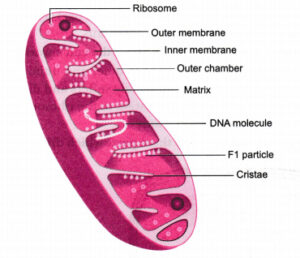
image credit: AmarCBSE
The DNA in eukaryotic cells is different to the DNA in prokaryotic cells because it coiled at regular intervals around cylinder-shaped proteins called histones. Prokaryotic DNA is not coiled around histones and exists as a simple loop of circular DNA.
How to draw a nucleus
A Eukaryotic nucleus should be drawn using two lines, with spaces for nuclear pores.
A2.2.7 Processes of life in living organisms
LAB: HUNTING FOR UNICELLULAR ORGANISMS (instructions in hard copy)
Unicelullar organisms are unique in that all the functions of life need to take place in one cell. Multicellular organisms may delegate specialised tissues or organs to carry out certain important life processes.
Universal life processes:
- Metabolism – biochemical reactions that sustain life (respiration is the best example)
- Reproduction – producing other members of the same species
- Growth – an increase in size or number of cells
- Homeostatis – regulating the internal environment of a body or cell
- Excretion – removal of waste products from cells or tissues
- Nutrition – consuming the nutrients required to sustain life (oxygen is an example in aerobically respiring organisms).
Q) How do paramecium achieve the universal life processes outlined above?
A2.2.8 Differences in Eukaryotic cell structure between animals, fungi and plant cells
Eukaryotic cell include animal, plant and fungal cells as well as unicellular organisms.
They are quite different from each other in some key feature.
Task: Study and observe the following images of a representative plant cell, animal cell and fungi
Previous
Next
Task: Discuss differences in the three kinds of cells. Write down 5 key differences.
Differences should include:
- Cell wall. Never present in animal cells. In fungi it is made from chitin, in plant cells from cellulose
- Plastids (an organelle with either pigment or food). Never present in animals or fungi, in plants they are common)
- Vacuole. In animals they are temporary, large permanent vacuoles are common in plants and fungi and may be used for storage
- Centrioles (used in cell division). Usually only found in animals.
- Cilia/flagella (hair like structures that generate movement). Found in animal cells (ex. sperm cell tails). Plant cells and fungi only have them if they have mobile male gametes (such as moss/bryophytes).
A2.2.9 Atypical cell structure in eukaryotes
Living tissue is normally formed from cells, each one with a distinct nucleus.
Some tissues are exceptions:
- Red blood cells have no nucleus at all. They exist simply to carry oxygen, die after a month, they are continously replaced by our bone marrow.
- Skeletal muscle is multinucleate (has lots of nuclei), and is made from long muscle fibres formed from cells that have fused together.
- Phloem sieve tubes (in plants). Are long tubes of cytoplasm used for transport in plants, they are formed from cells that breakdown to form tubes, the remains of the original cell boundaries opened by pores to form a tube with sieve like borders.
- Aseptate fungal hyphae (threadlike structures). Some fungi are made of threadlike structures which are not divided into cells by the boundaries called septa which other fungi typically have. These hyphae are multinucleate.
Previous
Next
Task
- A) Study the scanned diagrams of a plant and animal cell, taking notes regarding the functions of specific plant and animal cell organelles.
- B) Identify the specific organelles of a plant and animal cell from the scanned electron microscope images, annotating each label with notes on their specific functions.